タイトル厚生労働科学研究費補助金(難治性疾患克服研究事業)「Menkes 病・occipital horn 症候群の実態調査、早期診断基準確立、治療法開発に関する研究」平成23-24年度 総合研究報告書
- ページタイトル
- 厚生労働科学研究費補助金(難治性疾患克服研究事業)「Menkes 病・occipital horn 症候群の実態調査、早期診断基準確立、治療法開発に関する研究」平成23-24年度 総合研究報告書
76ページ中、13ページ目の概要を表示しています。
76ページ中、13ページ目の概要を表示しています。
厚生労働科学研究費補助金(難治性疾患克服研究事業)「Menkes 病・occipital horn 症候群の実態調査、早期診断基準確立、治療法開発に関する研究」平成23-24年度 総合研究報告書
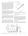
106 W. Bhadhprasit et al. / Journal of Trace Elements in Medicine and Biology 26 (2012) 105?108chloride solution (50 ?g of CuCl 2) on postnatal day 4, becausemacular mice die without this treatment. All mice were maintainedunder standard conditions. Macular mice were separatedinto control and treated groups. The latter group was treated witha subcutaneous injection of CuCl 2 (10 ?g) and oral administrationof disulfiram (0.3 mg/g body weight) twice a week from the age of7 days to 8 weeks, and then sacrificed. Control mice were given asubcutaneous injection of CuCl 2 (10 ?g) as above, but disulfiramwas replaced with double distilled water. Normal littermates wereused as normal controls. Body weights were measured twice a weekduring the treatment period. The cerebrum, cerebellum, kidney,liver, and intestines were dissected. Sera and tissues were stored at?80 ? C until analysis. This study was approved by Teikyo UniversitySchool of Medicine Animal Ethics Committee (07-035).Weight (g)252015105Control macularmiceTreated macularmice**Measurement of copper concentrationTissue samples were dried at 120 ? C for 12 h and wet-digestedwith concentrated HNO 3 by heating at 120 ? C, and the resultantresidues were dissolved in 2 mol/L HNO 3. Serum samples werewet-digested with concentrated HNO 3 by heating at 120 ? C. Copperconcentration was analyzed with a Hitachi Z-8100 atomic absorptionspectrophotometer (Hitachi Industries, Japan). All glasswarewas washed with nitric acid to avoid metal contamination.Assay for cytochrome c oxidase activityMitochondria were isolated from tissue samples immediatelyafter sacrifice using the Mitochondria Isolation Kit for Tissue(Pierce, Rockford, IL). Cytochrome c oxidase activity in the mitochondrialsolution was determined using the Cytochome c OxidaseAssay Kit (Sigma?Aldrich, St. Louis, MO). Protein concentration wasdetermined using the Pierce BCA Protein Assay Kit (Pierce, Rockford,IL).Measurement of catecholamine and enzyme concentrationsCerebrum and cerebellum were homogenized in 0.4 N perchloricacid and centrifuged at 4 ? C (12,000×g, 5 min). Catecholamines,including dopamine, noradrenaline, and adrenaline, in thesupernatant were analyzed using a catecholamine autoanalyzer(HLC-8030; Toso Ind., Tokyo, Japan). Serum levels of aspartateaminotransferase (AST), alanine aminotransferase (ALT), blood00 1 2 3 4 5 6 78Age (wks)Fig. 1. Body weight changes in control and treated macular mice from birth to 8weeks of age. Weights are presented as mean (SE) (control macular mice: n = 19;treated macular mice: n = 24). *p < 0.05.urea nitrogen (BUN), and creatinine were analyzed by OrientalYeast Co., Ltd. (Shiga, Japan).Statistical analysisData are presented as mean±standard error (SE). The SE is computedfrom known sample statistics, and it provides an unbiasedestimate of the standard deviation of the statistic. Differences inbody weight changes between the two groups were analyzed usingtwo-way repeated measure ANOVA. Differences in copper concentration,CCO activity and catecholamine between the two groupswere analyzed using one-way repeated measure ANOVA (Tukeypost hoc test); p < 0.05 was considered significant.Results and discussionAs shown in Fig. 1, the weight gain in treated macular micewas significantly higher than that of control macular mice afterweek 7. Fig. 2 shows the copper concentration in tissues and serum.Fig. 2. Copper concentrations in the cerebrum, cerebellum, liver, intestines, kidney, and serum of normal mice (n = 5), control macular mice (n = 19), and treated macularmice (n = 24). *p < 0.05; **p < 0.01.11