タイトル厚生労働科学研究費補助金(難治性疾患克服研究事業)「Menkes 病・occipital horn 症候群の実態調査、早期診断基準確立、治療法開発に関する研究」平成23-24年度 総合研究報告書
- ページタイトル
- 厚生労働科学研究費補助金(難治性疾患克服研究事業)「Menkes 病・occipital horn 症候群の実態調査、早期診断基準確立、治療法開発に関する研究」平成23-24年度 総合研究報告書
76ページ中、20ページ目の概要を表示しています。
76ページ中、20ページ目の概要を表示しています。
厚生労働科学研究費補助金(難治性疾患克服研究事業)「Menkes 病・occipital horn 症候群の実態調査、早期診断基準確立、治療法開発に関する研究」平成23-24年度 総合研究報告書
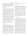
244 H. Kodama et al. / Brain & Development 33 (2011) 243?251to an excess of copper. The reason for this difference isrelated to the particular cell types in which the ATP7Aand ATP7B proteins are expressed. ATP7A is expressedin almost all cells with the exception of hepatocytes,whereas ATP7B is primarily expressed in hepatocytes.Here we review MD, OHS and WD with a focus on neurologicaspects.2. Copper homeostasisThe average daily intake of copper in healthy adultsis about 2 mg. Ingested copper is absorbed from theintestine into the blood, and the majority is then transportedinto the liver. The majority of copper in the liveris excreted via the bile, but a small amount is excreted inthe urine. Fig. 1 shows the molecular mechanism of coppermetabolism in cells. Ctr1 is a high-affinity coppertransporter located on the plasma membrane of the cellsand mediates copper uptake. Copper in the cytosol isdelivered to Cu/Zn superoxide dismutase in the cytosol,to the Golgi apparatus, and to mitochondria by Ccs2,HAH1 (ATOX1) and Cox 17, respectively, and aregenerically named copper chaperones [1]. ATP7A andATP7B are localized to the trans-Golgi membrane andtransport copper from the cytosol into the Golgi apparatuswithin cells. Copper transported into the Golgiapparatus is excreted from cells as a part of copperenzymes. ATP7A is expressed in almost all cells otherthan hepatocytes, including those of the intestine, kidneyand components of the blood brain barrier; ATP7Bis mainly expressed in hepatocytes and acts to excretecopper into the bile and blood [2,3].Genetic disorders of copper metabolism in humansmanifest in the form of MD, OHS and WD (Table 1).3. Menkes disease (MD) and occipital horn syndrome(OHS)3.1. GeneticsThe phenotypic features of ATP7A mutations can bedivided in at least three categories; Classical MD withdeath in the early childhood (generally called as MD),mild MD with long survival, and OHS (the mildest features)[4]. Inheritance of MD and OHS is X-linked recessive;patients are typically male, and their mothers areheterozygous carriers of the disease. The incidence ofMD in Japan is estimated to be 1/140,000 live malebirths [5]. A small number of females with X-linkedchromosomal abnormalities have also been reported tobe affected by MD [1]. Patients with MD exhibit a largevariety of mutations in the ATP7A gene [1,6,7]. Mollerdescribed that they had identified about 357 differentmutations [4].OHS and mild MD are extremely rare. Major mutationsin the ATP7A gene in OHS and mild MD aresplice-site or missense mutations [4], and residual activityof ATP7A still exists [6,7].3.2. PathologyIn MD-affected cells, copper accumulates in the cytosoland cannot be excreted. Copper accumulation in theintestine results in a failure of copper absorption, whichleads to copper deficiency in the body and reduces theactivity of copper-dependent enzymes. Copper alsoaccumulates in the components of the blood?brain barrierand cannot be transported from the blood vessels toneurons. The characteristic features of MD can beexplained by a decrease in the activity of copper-dependentenzymes (Table 2).Neurologic degeneration in MD is mainly caused bydecreased activity of cytochrome C oxidase in neurons.In addition, subdural hemorrhage often occurs secondaryto disorders of brain arteries due to decreased activityof lysyl oxidase, resulting in neurologic damage.Hypotonia may be caused by reduced activity of cytochromeC oxidase in the muscle [8].Characteristics of OHS include connective tissue disorderscaused by a decrease in lysyl oxidase activity.3.3. Clinical features3.3.1. Neurologic manifestationsCharacteristic clinical features including seizures,delayed development, marked muscular hypotonia andabnormal hair, become prominent between the ages of2 and 4 months when copper deficiency becomesadvanced. Because clinical abnormalities are absent orsubtle in affected newborns, the diagnosis is difficultprior to 2 months of age. As the disease progresses,patients are bedridden and never smile. Most patientswith MD die by the age of 3 years, although somepatients survive into their teenage years [1,7].Epilepsy is observed in most patients with MD.Bahi-Buisson et al. reported the characteristics of epilepsyas divided into three periods [9]. Focal clonicstatus epilepticus and intractable infantile spasms areobserved in the early stage (median age: 3 months)and intermediate stage (median age: 10 months),respectively. Multifocal seizures, tonic spasms andmyoclonus are observed in the late stage (medianage: 25 months). Ozawa et al. also reported that infantilespasms with EEGs containing hypsarrhythmic patternsare observed in 50% of epileptic patients withMD [10]. Brain MRIs of the patient shows brain atrophyand delayed myelination or demyelination. Subduralhemorrhage/effusion is often observed (Fig. 2).Magnetic resonance angiography shows a tortuosityof intracranial and cervical blood vessels [1,7].1Additionally, H-magnetic resonance spectroscopy( 1 H-MRS) shows a lactate peak and a decrease in18