タイトル厚生労働科学研究費補助金(難治性疾患克服研究事業)「Menkes 病・occipital horn 症候群の実態調査、早期診断基準確立、治療法開発に関する研究」平成23-24年度 総合研究報告書
- ページタイトル
- 厚生労働科学研究費補助金(難治性疾患克服研究事業)「Menkes 病・occipital horn 症候群の実態調査、早期診断基準確立、治療法開発に関する研究」平成23-24年度 総合研究報告書
76ページ中、25ページ目の概要を表示しています。
76ページ中、25ページ目の概要を表示しています。
厚生労働科学研究費補助金(難治性疾患克服研究事業)「Menkes 病・occipital horn 症候群の実態調査、早期診断基準確立、治療法開発に関する研究」平成23-24年度 総合研究報告書
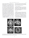
H. Kodama et al. / Brain & Development 33 (2011) 243?251 249the most common manifestation is dysarthria (57.6%),followed by dystonia (42.4%), parkinsonism (17.3%),chorea or athetosis (15.3%), and seizures (4.7%). Psychiatricmanifestations may be present as often as 30?50%of the time prior to a diagnosis of WD [23]. Seizuresoccur with a prevalence of 4?8.3% and sometimes havebeen associated with de-coppering therapy [24]. Thetypes of seizures are variable and include generalizedtonic?clonic, simple partial, complex partial, and partialseizures with secondary generalized periodic myoclonus[24]. Status epilepticus is rare. Pestana-Knight et al.reported a patient with WD who developed status epilepticusduring therapy with tetrathiomolybdate; the seizureswere controlled with fosphenytoin, midazolam,and levetiracetam [25]. Early diagnosis and prompt initiationof treatment is very important for patients withneurologic changes because treatment outcome worsenswith a delayed initiation of therapy. Copper levels in thecerebrospinal fluid increase in patients with neurologicsymptoms, and the levels decrease to within normalrange after treatment, suggesting that the copper levelis a useful marker for monitoring therapy in patientswith neurologic symptoms [26].On MRI scan, a high signal on T2 and low signal onT 1 imaging are observed in the lentiform and caudatenuclei, thalamus, brainstem and white matter. High signalT1 images, like those in portal-systemic encephalopathy,are also observed [27]. Loss of cerebral whitematter has rarely been reported (Fig. 6).4.3.2. Other manifestationsHepatic symptoms demonstrate a broad spectrum ofchronic hepatitis, acute hepatitis, cirrhosis and fulminanthepatic failure. In addition, initial symptoms suchas hematuria, anemia, arthritis, hemolysis, cardiomyopathy,dysrhythmias and hypersalivation are variable andmake early diagnosis difficult [1].4.4. DiagnosisDiagnosis is usually made by detecting low serum levelsof copper and ceruloplasmin (<20 mg/dl by immunoassay),high copper concentrations in the liver (>250 lg/g dry weight), high copper excretion in the urine(>100 lg/day), and a penicillamine challenge test (urinarycopper excretion > 1600 lg/day). Although aDNA-based diagnosis is also available, approximately17% of patients diagnosed with WD on the basis of clinicalsymptoms and biochemical data have no mutationsin the coding regions of ATP7B [18]. Scoring systems forFig. 6. MRI of patients with WD. T2-weighted images show a high signal in the capsula externa, thalamus and basal ganglia (a,b).“Face of GiantPanda”finding appears due to high signal in the midbrain on T2-weighted imaging (c) [36] (used with permission). Loss of left frontal cerebral whitematter is observed in a neurologic patient with WD who suffered from right hemiplegia.23