タイトル厚生労働科学研究費補助金(難治性疾患克服研究事業)「Menkes 病・occipital horn 症候群の実態調査、早期診断基準確立、治療法開発に関する研究」平成23-24年度 総合研究報告書
- ページタイトル
- 厚生労働科学研究費補助金(難治性疾患克服研究事業)「Menkes 病・occipital horn 症候群の実態調査、早期診断基準確立、治療法開発に関する研究」平成23-24年度 総合研究報告書
76ページ中、33ページ目の概要を表示しています。
76ページ中、33ページ目の概要を表示しています。
厚生労働科学研究費補助金(難治性疾患克服研究事業)「Menkes 病・occipital horn 症候群の実態調査、早期診断基準確立、治療法開発に関する研究」平成23-24年度 総合研究報告書
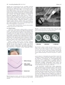
242 Current Drug Metabolism, 2012, Vol. 13, No. 3 Kodama et al.apparatus and is secreted from the cells. Accordingly, parenteraladministration of copper-histidine cannot improve enzyme activitiesbecause the administered copper is not transported into theGolgi apparatus due to ATP7A defects. In fact, serum and urinelevels of bone metabolic markers are poorly improved by copperhistidinetherapy in patients with MD [49]. Neurochemical patternsin the serum and cerebrospinal fluid of patients with MD resembledthat of patients with congenital deficiencies of dopamine ?-hydroxylase, suggesting that this enzyme activity is reduced inpatients with MD [50]. Dopamine s-hydroxylase is also a secretoryenzyme, and thus its enzyme activity could not be increased by acopper-histidine injection. Another characteristic feature of MD issevere muscular hypotonia. Although the pathology of muscularhypotonia remains unknown, reduced activity of cytochrome Coxidase in muscles may be involved [51]. In the kidneys of macularmice, copper accumulates in the cytosol of proximal tubular cells,but not in the distal tubules or glomeruli [44] .In contrast, OHS is characterized by connective tissue disorderscaused by decreased lysyl oxidase activity.8r4.3. Clinical FeaturesCharacteristic clinical features of MD and OHS are summarizedin Tables 1 and 2, and are shown in Figs. 5-9. Developmental delay,seizures, and marked muscular hypotonia become prominent aftertwo months of age when copper deficiency is advanced. Diagnosisis difficult prior to two months of age because clinical abnormalitiesare subtle or sometimes absent in affected newborns [52]. Neurodegenerationand connective tissue abnormalities do not improveand progress when copper-histidine therapy is initiated at 2 monthsof age or older. As the disease progresses, patients become bedriddenand are unable to smile or speak. Although most patients die bythe age of three, a few survive beyond 20 years of age [40,52].Epilepsy, including infantile spasms, myoclonus, multifocalseizures, and tonic spasms, are observed in over 90% of patientswith MD who have been treated after 2 months of age [53,54].Magnetic resonance imaging (MRI) reveals brain atrophy and delayedmyelination or demyelination, and subdural hemorrhage isoften observed (Fig. 7). Magnetic resonance angiography (MRA)aFig. (6). A 2 year-old patient with Menkes disease treated with copperhistidineinjections since the age of 8 months. Despite treatment, he suffersfrom severe muscle hypotonia and cannot hold up his head.2 Months 8 Months 11 MonthsFig. (7). Brain CT images of a patient with Menkes disease at 2, 8, and 11months of age. The image was taken at the age of 2 months because of ahead injury. This was prior to diagnosis of MD as no neurological symptomswere observed at that time. The patient was diagnosed with MD at theage of 8 months, with brain atrophy progressing despite copper-histidinetreatment. Subdural hemorrhage was observed in the patient at 11 months ofage.b2 years oldBefore therapyAfter copperhistidinetherapyNormal hairFig. (5). Depigmented, lusterless, and kinky hair in a 3-month-old patientwith Menkes disease. Hair abnormalities were improved by copper-histidineinjections.reveals tortuosity of intracranial and cervical blood vessels [16].1H-magnetic resonance spectroscopy (MRS) shows a lactate peakand decreased N-acetylaspartate and creatinine/phosphocreatinelevels [55]. Lesions of hypointensity on T1-weighted images andhyperintensity on T2-weighted images are transiently observed intemporal lobes, and appear similar to stroke-like lesions observed inmitochondrial myopathy, encephalopathy, lactate acidosis, andstroke-like episodes (MELAS). This suggests that the lesions observedin MD may be due to ischemic events [56].Hair abnormalities, including kinky, tangled, depigmented,friable, and sparse hair, are characteristic features of MD and oftendiagnostic (Fig. 5). Bladder diverticula, osteoporosis, skin and jointlaxity, and arterial abnormalities are connective tissue changescaused by decreased lysyl oxidase activity. Patients with MD haveintractable and chronic diarrhea that results in severe malnutrition;however, the etiology is unclear. Urinary infection is common andmost likely due to bladder diverticula. Although severe copper toxicityis not typically observed, urinary s2-microglobulin levels areelevated in patients, suggesting that toxicity does occur in renalproximal tubules [57].31