タイトル厚生労働科学研究費補助金(難治性疾患克服研究事業)「Menkes 病・occipital horn 症候群の実態調査、早期診断基準確立、治療法開発に関する研究」平成23-24年度 総合研究報告書
- ページタイトル
- 厚生労働科学研究費補助金(難治性疾患克服研究事業)「Menkes 病・occipital horn 症候群の実態調査、早期診断基準確立、治療法開発に関する研究」平成23-24年度 総合研究報告書
76ページ中、35ページ目の概要を表示しています。
76ページ中、35ページ目の概要を表示しています。
厚生労働科学研究費補助金(難治性疾患克服研究事業)「Menkes 病・occipital horn 症候群の実態調査、早期診断基準確立、治療法開発に関する研究」平成23-24年度 総合研究報告書
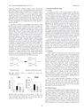
244 Current Drug Metabolism, 2012, Vol. 13, No. 3 Kodama et al.with known mutations resulting in partial ATP7A function hadneither clinical seizures nor electroencephalographic abnormalities[65]. These findings suggest that differences in treatment responsewould also depend on residual ATP7A activity. Symptoms relatingto connective tissue disorders are scarcely improved by copperhistidinetreatment. This is explained by the fact that the administeredcopper cannot be transported from the cytosol into the Golgiapparatus where it is incorporated into lysyl oxidase. Patients withMD who are diagnosed and treated early show phenotypic featuresof OHS. Unfortunately, no treatment trials have been reported inpatients with OHS.An effective treatment for neurological and connective tissuedisorders has not yet been established. If the delivery of copper intothe trans-Golgi apparatus of affected cells could be achieved, thencopper treatment would probably normalize the activity of lysyloxidase and improve connective tissue disorders associated withMD and OHS. Likewise, if copper could be delivered to the Golgiapparatus within cells comprising the blood-brain barrier, copperwould reach neurons and be incorporated into cuproenzymes includingcytochrome C oxidase in the neurons. We previously reportedthat combination therapy with copper and diethyldithiocarbamate(DEDTC, Fig. 10), a lypophilic chelator, improves copperconcentration, cytochrome C oxidase activity, and catecholaminemetabolism in the brains of macular mice (Fig. 11) [66]. Takeda etal reported a 3-year-old patient treated with copper-histidine andoral disulfiram (DEDTC dimmer) for a period of 2 years [67]. Serumcopper and ceruloplasmin levels increased and were higherthan those when patients were administered copper-histidine alone.In addition, we observed a smile from the patient administered thecombination therapy. The hydrophobicity of DEDTC seems to supportpassage of copper chelated with this compound through themembrane. To establish the utility of this therapy, further studiesfocusing on survival, biochemical parameters, and clinical outcomein both animal models and patients with MD are necessary.Fig. (10). Chemical reaction of chelation by sodium N,Ndiethyldithiocarbamate.ng/mg wet weightDiethyldithiocarbonate543210Cu***8 5 6 3CC MA MB MCdiethyldithiocarbonate-copper complex?mol/min/mg protein250200150100Fig. (11). Copper concentrations (Cu) and cytochrome C oxidase (CCO)activity in the cerebrum of macular mice. MA, macular mice treated withcopper and DEDTC; MB, macular mice treated with copper only; MC,macular mice without treatment. (*p<0.05; ** p<0.01.500CCO*8 7 6 3CC MA MB MC*V. WILSON’S DISEASE (WD)5.1. GeneticsThe global prevalence of WD is approximately 1/30,000 newborns,although this varies across populations [68]. This autosomalrecessive disorder is caused by mutations in the ATP7B gene, andover 480 mutations have been reported (http//www.medgen.med.ualberta.ca/database). The R778L substitution is the most commonmutation occurring in Asian patients, while the H1069Q mutation ismostly seen in European patients [17,69,70]. A correlation betweengenotype and phenotype has not been found in patients with WD,although several mutations correlate well with an early onset of thedisease [71,72]. WD manifestations may be influenced by genevariants of baculoviral IAP repeat-containing protein 4/X-linkedinhibitor of apoptosis protein (BIRC4/XIAP), which is antiapoptoticand likely acts as a regulator of copper-induced cell death[73]. Gupta et al recently reported that a 9-year-old and 6-montholdpatient with high neurological predominance and mild hepaticsymptoms not only had heterozygous mutations in ATP7B, but alsohad mutations in COMMD1 [74]. The authors, however, concludedfrom a genetic analysis of 108 patients that COMMD1 variants donot contribute to the phenotypic heterogeneity observed in WD.Two animal models have been reported for WD. Long-EvansCinnamon (LEC) rats harbor a deletion in the ATP7B gene, accumulatelarge amounts of copper in the liver, and develop chronichepatitis, which eventually leads to hepatocellular carcinoma[75,76]. Toxic milk mice have a mutation in the transmembranedomain of atp7b, and show decreased levels of ceruloplasmin withaccumulation of copper in the liver, which eventually leads to cirrhosis[77].5.2. PathologyCopper cannot be transported from the cytosol into the Golgiapparatus, where copper is incorporated into apo-ceruloplasmin inhepatocytes of patients with WD. Accordingly, secretion of copperas holo-ceruloplasmin into the blood is affected. Excretion of copperinto bile is also affected (Fig 3b), resulting in copper accumulationin the liver. During the early stage of the disease, copper isdiffusely distributed as metallothionein-copper in the hepatocyticcytosol. With disease progression, copper accumulates in thelysosomes. This excess copper induces free radical production,which causes cellular damage via oxidative stress. Furthermore,serum levels of ceruloplasmin decrease, at which point ceruloplasmin-boundcopper decreases in the serum. Excess copper in theliver is released into the plasma as non-ceruloplasmin-bound copper,i.e., copper bound to albumin or amino acids, although therelease mechanism is unclear [78]. The increase in serum nonceruloplasmin-boundcopper results in elevated urinary copper excretionand copper deposition in various tissues, including the brain,kidney, cornea, muscle, bone, and joint [17].Iron as well as copper reportedly accumulates in the liver ofpatients with WD [79]. Due to the oxidase activity in ceruloplasmin,which converts ferrous iron to ferric iron, decreased ceruloplasminlevels in WD disrupt iron homeostasis [80]. Liver damagein patients with WD may be caused, in part, by iron accumulation,which is also toxic to the liver [79].5.3. Clinical FeaturesProminent clinical features of WD include hepatic and neurological/psychiatricsymptoms. Hepatic symptoms range from acuteand chronic hepatitis to cirrhosis and fulminant hepatic failure.Although serum levels of transaminases are high in infants withWD, hepatic disorders usually occur after 8 years of age. Neurologicalsymptoms appear after 12 years of age and are characterizedby extrapyramidal effects, which include dysarthria, dystonia,tremor, choreoathetosis, and ataxia [81]. Cognitive impairment anddepression are also common in patients with WD. Seizures occurwith a prevalence of 4-8.3% and are sometimes associated with de-33